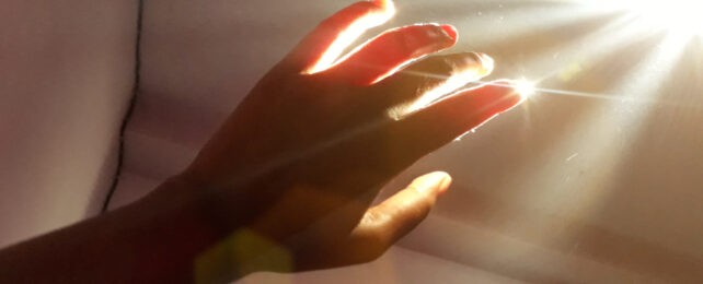
We Glow While Alive: A Visible Light Emitted by Living Cells Vanishes at Death

A team from the University of Calgary and the National Research Council of Canada reports ultraweak photon emission (UPE) in living organisms, detectable in whole mice and in certain plant leaves. In controlled experiments, researchers captured photons in the visible range from subjects while they were alive and after death. The post-death signal dropped sharply, suggesting the glow fades with death. While the idea that living tissue emits light may sound fringe, the work hints at a potential non-invasive way to gauge health, though the science remains controversial and far from settled.
In This Article:
- What are biophotons? A faint glow from living tissue, long debated
- The live-versus-dead experiment: four mice in a dark box, then euthanized
- Plants also glow when stressed: Arabidopsis thaliana and a dwarf umbrella tree reveal a stress-light link
- Implications and cautions: could a glow become a diagnostic tool?
What are biophotons? A faint glow from living tissue, long debated
Biophotons are light produced by biological processes, typically in the 200–1,000 nanometer range. The signals are extraordinarily faint and are easily overwhelmed by ambient light and heat, making robust detection challenging. Researchers point to reactive oxygen species generated under stress as a plausible source of these emissions, transforming energy into photons as molecules rearrange.
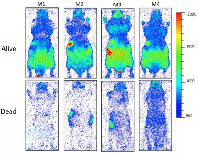
The live-versus-dead experiment: four mice in a dark box, then euthanized
To test whether the glow scales to whole organisms, four immobilized mice were placed in a dark box and imaged for an hour while alive, then imaged for another hour after they were euthanized. The animals were warmed to body temperature to keep heat from being a confounding variable. Using electron-multiplying CCD and CCD cameras, the team captured individual photons in the visible band emitted by tissues before and after death, with a clear drop in intensity after euthanasia.
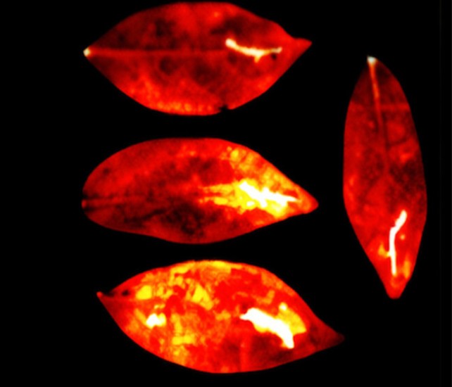
Plants also glow when stressed: Arabidopsis thaliana and a dwarf umbrella tree reveal a stress-light link
In parallel, researchers examined leaves from Arabidopsis thaliana and the dwarf umbrella tree (Heptapleurum arboricola), applying physical injuries and chemical stress. Across 16 hours of imaging, the injured parts of all leaves remained brighter than the uninjured parts, supporting a link between stress and light emission driven by reactive oxygen species. The researchers state: "Our results show that the injury parts in all leaves were significantly brighter than the uninjured parts of the leaves during all 16 hours of imaging."
Implications and cautions: could a glow become a diagnostic tool?
If such faint biophotonic glow tracks cellular health, it could someday become a non-invasive diagnostic tool for humans, crops, or biological samples. Yet the signal is extremely weak and easily swamped by ambient light; results require replication and careful scrutiny before any practical use. The findings were published in The Journal of Physical Chemistry Letters, with an earlier version of the article first published in May 2025.